


Ultrasound imaging is the technique of using high-frequency sound waves to penetrate body tissues and produce a picture of the tissues inside a patient. This is the same principle that is used to produce undersea images using sonar. An ultrasound image is produced by measuring the time required for sound waves to leave and return to the ultrasound probe from various tissues in the body. A computer in the ultrasound machine converts this information into an image on the monitor.
We are able to use ultrasound to examine many organs that are poorly visualized by radiography and to observe the actual motions of certain organs, most particularly the heart. Ultrasonography allows the operator to discover cysts and tumors within organs such as kidneys, liver, spleen and urinary bladder, and to find abnormalities of other abdominal organs that might otherwise only be found by surgery or more expensive imaging modalities, such as CT or MRI scans. Heart function and heart valve abnormalities may also be seen and measured using ultrasonography.
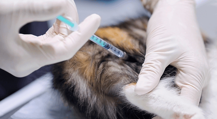
Our veterinarians are happy to be able to provide these ultrasound services to our St. Petersburg and Tampa Bay Area clients and their pets.
Ultrasound is particularly effective at looking inside solid or fluid-filled structures, such as those in the abdomen. This is an area where radiography (x-ray), the traditional imaging mode, is limited by similarities in density of adjacent organs. For example, where radiography produces a shadow of the outline of the liver, ultrasound shows the various blood vessels and ducts within the liver, gives clues as to the organ’s texture, and even demonstrates masses within the organ.
On the other hand, ultrasound is a poor choice for imaging bones or lung tissue. High-density structures such as bone, and very low-density materials, such as air, completely block ultrasound wave transmission. Fortunately, these are the very areas in which radiographic images excel. As a result, having access to both radiography and ultrasound gives the veterinarian the ability to image almost all body areas effectively.
Radiology
In the last 20 years, medical imaging technologies have allowed veterinarians to see inside animals in different ways. Ultrasound uses sound waves, magnetic resonance imaging (MRI) uses the spin of hydrogen atoms, and computed tomography (CT) uses x-rays analyzed by a computer.
Despite these advances, diagnostic x-ray remains one of the most convenient, economical and versatile means of diagnostic imaging. Radiography can help detect many diseases throughout the body in dogs, cats, and exotic pets. Our staff is skilled in producing high-quality diagnostic x-ray images to aid our veterinarians in diagnosing a plethora of medical conditions. When needed, we also enlist the services of board-certified radiology specialists to aid in complex cases.
Our veterinarians have access to CT and MRI imaging through local, specialty referral practices when these less frequently used and more costly procedures are indicated.
“Veterinary Diagnostic Imaging” plays an important role in diagnosing and monitoring treatment in all species of animals, and we are happy to be able to provide this service to our St. Petersburg and Tampa Bay Area clients and their pets.
Please contact our veterinarians at Animal Medical Hospital in Saint Petersburg, Florida (FL) at 727-896-7127 for more information.